Introduction
The gastrointestinal tract (GIT) constitutes an organ system that is of critical importance for our health and well-being. Some of the major functions of the GI tract are related to the ingestion of food, digestion, absorption of nutrients and defecation.Additionally, the GIT mucosa contains the largest amount of immune cells in our body constantly monitoring the contents of the gut lumen aiming to prevent the entrance of hazardous or disease-causing microorganisms. Also, the gut lumen contains myriads of harmless or beneficial microorganisms, i.e. around 10 times the number of cells in our body, that take part in the digestive process, but above all are essential for the development and maintenance of a healthy immune system. Thus, the gut microflora, the intestinal epithelium and the intestinal mucosal immune system interact in a reciprocal fashion to maintain tissue homeostasis and vital body functions.
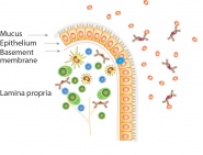
 Schematical drawing of a villus top from the small intestine with immune cells in the lamina propria and epithelial cells with microvilli, all covered with a protective mucus layer. Photo: Tor Lea
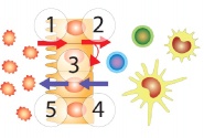
 The figure depicts the reciprocal regulatory interactions taking place between the gut microflora, the intestinal epithelium and different immune cells. Photo: Tor Lea
MAIN TOPICS UNDER SCRUTINY:
1
The significance of food and the gut flora for intestinal tissue homeostasis and the immunoregulatory properties of the intestinal epithelium
2
The inflammatory process and its significance for intestinal tissue homeostasis
3
Stem cells in intestinal tissue homeostasis
POSTER: "Identification and localization of human intestinal stem cells (hISCs)"
4
Intestinal tissue homeostasis and inflammatory bowel disease (IBD)
5
Intestinal tissue homeostasis, the epithelial-mesenchymal transition (EMT) and colon cancer
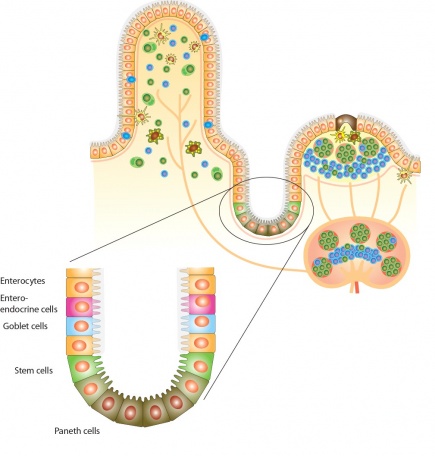
 The figure shows the structure of an intestinal villus and a crypt with associated secondary lymphoid tissue in the form of a Peyers patch and a mesenteric lymph node. Enlarged are the different cell types of the intestinal crypt epithelium with Paneth cells producing anti-microbial peptides, stem cells, mucin-producing goblet cells, enteroendocrine cells producing hormon-like compounds and the adsorptive enterocytes. Photo: Tor Lea